Research Stories
World's first development of ultrahigh temporal resolution brain neuronal activity imaging technology that can see...
[ Published in the international academic journal 'Science' ]
Objective and quantitative evaluation of cognitive impairment or mental disorders in degenerative brain disease
Biomedical Engineering
Prof.
PARK, JANGYEON
World's first development of ultrahigh temporal resolution
brain neuronal activity imaging technology that can see the flow of thoughts
The research team led by Professor Jang-Yeon Park of the Department of Biomedical Engineering at Sungkyunkwan University has developed the world’s first next-generation brain function imaging technology that can directly image brain neuronal activity in vivo with ultra-high resolution of a few milliseconds. The main verification of the proposed brain neuronal activity imaging technology was carried out in collaboration with the research team of Professor Jeehyeon Kwag of Korea University (currently Seoul National University), an electrophysiology research group.
Non-invasive brain function imaging (or neuroimaging) techniques play an important role in elucidating how the brain functions in vivo. However, the most widely used non-invasive brain functional imaging techniques, such as electroencephalogram(EEG), magnetoencephalogram(MEG), and functional magnetic resonance imaging(fMRI), have distinct advantages and disadvantages in terms of temporal and spatial resolution, acting as important limitations for in vivo brain research. For example, EEG and MEG give low spatial resolution (~centimeters, cm) despite high temporal resolution (~milliseconds, ms), and functional magnetic resonance imaging (fMRI) provides low temporal resolution (~seconds, sec) despite high spatial resolution (~millimeters, mm), providing only indirect neural activity information based on blood flow.
Prof. Park’s research team predicted that direct imaging of neural activity (DIANA) would be possible if magnetic resonance imaging (MRI) with a temporal resolution of several milliseconds, which is comparable to the time scale of neural action potentials, was implemented. The research team realized ultra-high time resolution in milliseconds using a method of segmenting image data. Using this, the neural activity directly transmitted in the brain neural network could also be imaged. In addition, the research team proposed an important biophysical hypothesis for possible signal sources of DIANA.
Prof. Par’s research team verified the proposed brain neuronal activity imaging technique using an in vivo mouse brain in a 9.4T animal MRI system. By applying repetitive electrical stimulation to the mouse whiskers, time series images of DIANA in neural networks including the thalamus and primary somatosensory cortex (S1) were acquired with s spatial resolution of 0.22 mm and a temporal resolution of 5 ms. As a result, DIANA responses in S1 were confirmed at 20-25 ms and at 10-15 ms in the thalamus, which successfully imaged how neuronal activity propagates in the thalamus-cortical pathway. In addition, the research team proposed a change in T2 relaxation time due to changes in membrane potential and cell volume accompanying neural activity as a new contrast mechanism for DIANA.
The novel brain function imaging technique proposed by Prof. Park’s research team can show how neural activity is transmitted in the brain neural network in vivo, along with direct imaging of neural activity, by imaging brain neural activity with a time resolution of a few milliseconds. Through this next-generation brain function imaging technology, it is expected to implement a dynamic brain neural network model close to reality that can reflect and express how brain functions are actually performed in various cognitive processes. From a clinical point of view, it is also expected to contribute greatly to individual precision diagnosis, the trend of modern medicine, by enabling objective and quantitative evaluation of cognitive impairments in neurodegenerative diseases such as Alzheimer’s disease and Parkinson’s disease, as well as mental disorders including depression and neurodevelopmental disorders.
Professor Park said, “This study is very meaningful in that it realized an in vivo brain neural activity imaging with both high temporal and high spatial resolution, which has been a long-cherished dream in the field of brain function imaging.”, and also said, “In particular, being able to image neural activity and its propagation in high spatiotemporal resolution in the neural network means that the flow of information, that is, the flow of thoughts, can be seen in the cognitive process in the brain neural network. Through this, it is expected that an in-depth understanding of the ‘thinking brain’ will be possible by elucidating the hierarchical connectivity of brain functions.” He finally added, “If it is proven that DIANA can also be applied to humans, it could be a game changer in the field of brain science.”
The results of this study were published as a Research Article on October 14, 2022 in ‘Science’ (IF: 47.728), along with Perspectives, a commentary article published together with noteworthy papers. Also, along with the publication of the paper, Nature news covered an article on this paper. In addition, The Scientist(UK), STAT news(USA), and ChosunBiz(Korea) also published articles about this paper.
※ Paper Title: “In vivo direct imaging of neuronal activity at high temporo-spatial resolution”
※ Science, https://www.science.org/doi/10.1126/science.abh4340
[Fig. 1] High temporo-spatial resolution DIANA captures thalamocortical spike propagation.
(A) Illustration of the DIANA experiment to image contralateral S1BF and thalamus applying electrical stimulation to left whisker pad in an anesthetized mouse on a 9.4 T scanner (right) and brain imaging of a coronal slice containing both thalamus and S1BF regions (left). (B) BOLD activation map obtained as a reference (n = 10 mice). (C to E) Time series of t-value maps of DIANA for 30 ms after whisker-pad stimulation in 5 ms temporal resolution from 5 mice (C), percent changes in DIANA signals (D), and bar graphs showing the mean latencies of peak DIANA responses from the thalamus (green) and contralateral S1BF (magenta) (E) (n = 10 mice, ****: p < 0.0001, paired t-test). (F) Top: Illustration of electrophysiological recording in mice in vivo with silicon probes implanted in the thalamus and the contralateral S1BF applying electrical whisker-pad stimulation, Bottom: Electrode track marking using a fluorescent lipophilic dye (DiI). (G and H) Multi-unit activity (MUA) (black trace, top) from which single-unit spikes (bottom) were analyzed in the thalamus (green) (G) and the contralateral S1BF (magenta) (H). (I) Post-stimulus time histogram (PSTH) of the whisker-pad stimulation-responsive single units over time in the thalamus (top) and contralateral S1BF (bottom) with DIANA signals superimposed for comparison. (J) Bar graph showing the latencies of peak spike firing rates of whisker-pad stimulation-responsive single units recorded from the thalamus (light green, n = 23 units from 10 mice) and contralateral S1BF (light magenta, n = 23 units from 5 mice). Vertical dotted lines indicate the whisker-pad stimulation on a set time (red) (D, and G to I) and latency of peak spike firing rate (thalamus, green; contralatera lS1BF, magenta) (I). (****:p < 0.0001, unpaired t-test). All data are mean ± SEM.
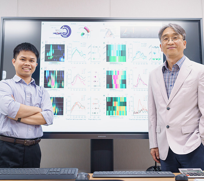
[Fig. 2] Sublayer-specific DIANA responses revealed functionally distinct sublayer-specific microcircuits.
(A and B) Illustration of DIANAexperiment (A) and in vivo spike recording (B) in VPMd, VPMv, POm, S1BF, and S2 in response to electrical whisker-pad stimulation. Yellow dotted boxes in (A, right) indicate extraction areas of DIANA heatmaps. (C) Heatmap (left) and temporal profile (middle) for percent change in DIANA signal, displayed with a mean latency of peak DIANA response from VPMd (dark green), VPMv (green), and POm (lightgreen) (right) (n = 9 mice). (D) Heatmap of in vivo-recorded single-unit spike firing rate normalized to the peak firing rate (left) and temporal profile of spike firing rate (middle), displayed with a mean latency of peak spike firing rate in VPMd (darkgreen, n = 22 units from 16 mice), VPMv (green, n = 15 units from 16 mice), and POm (lightgreen, n = 59 units from 14 mice) (right). (E and F) Same as (C) and (D) but for DIANA experiments (n = 9 mice) and spikes recorded from S1BF in L2/3 (light pink, n = 9 units from 28 mice), L4 (pink, n = 51 units from 28 mice), L5 (magenta, n = 60 units from 28 mice), and L6 (dark magenta, n = 18 units from 28 mice). (G and H) Same as (C) and (D) but for DIANA experiments (n = 9 mice) and spikes recorded from S2 in L2/3 (light orange, n = 6 units from 20 mice), L4 (orange,n = 27 units from 20 mice), L5 (brown, n = 35 units from 20 mice), and L6 (dark brown, n = 14 units from 20 mice). The base in DIANA heatmaps indicates the average of pre-stimulation frames. The asterisk in the middle of (C) and (E) indicates the statistically significant negative signal. All data are mean ± SEM. *: p < 0.05, **: p < 0.01, ****: p < 0.0001, n.s.: p > 0.05 for paired, unpaired, and Welch t-test.
* News Link
Nature
https://www.nature.com/articles/d41586-022-03276-5
TheScientist
https://www.the-scientist.com/news-opinion/new-mri-technique-tracks-brain-activity-at-millisecond-timescales-70626
STAT news
https://www.statnews.com/2022/10/13/faster-brain-imaging-seems-to-overcome-limitations-of-mri-scans/
Wangyi Newspaper


