Research Stories
Development of muscle-mimetic cell-laden Nanofiber using 3D Cell-Electrospinning
Enhancing muscle regeneration with anisotropically arranged nanofibers
Bio-Mechatronic Engineering
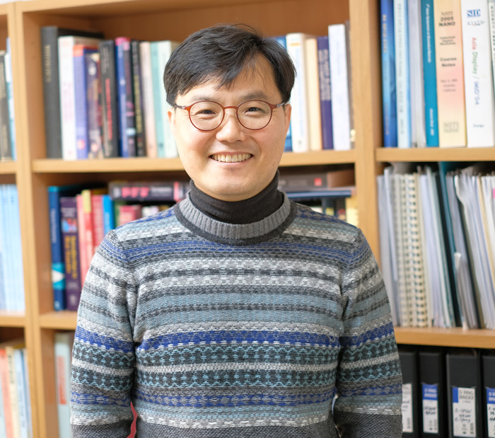
Prof.
KIM, GEUNHYUNG
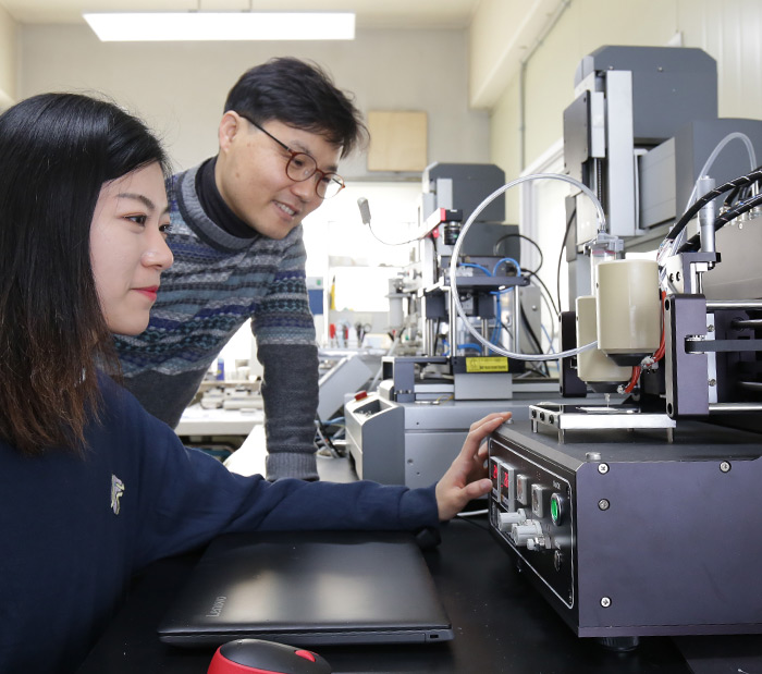
Prof. Geun Hyung KIM and his research team reported that they have successfully aligned the nanofibrous structure by producing live myoblast cells and bioink suitable for electrospinning. Nano-muscular fibers implanted with live myoblast cells acted as if it were a real muscle tissue and accelerated the regeneration of muscle tissue by guiding the muscle cell to grow in a uniaxial direction.
Tissue Regeneration Engineering is a field of study developed to improve the regeneration process of damaged tissues/organs by inserting a biological substitute, which is called scaffold. 3D cell-printing and electrospinning has been widely used for this process. However, the cells cultured by 3D cell-printing and electrospinning grew randomly, which was a serious problem for muscles that required its cells to be aligned for proper regeneration.
To control cell morphology, they have developed electrospinning to a cell-electrospinning process. The research team used a biocompatible hydrogel to generate cell-laden nanofibers. Also, the hydrogel was added with a material with high processability to produce a bioink, which was applied with high-voltage direct current (Figure 1). After this, myoblast-laden nanofiber can be generated with an aligned pattern.
-the myoblast-laden nanofibers showed over 90% of initial cell viability, which was a sign that it overcame the problem of low cell viability from the previous conventional cell-electrospinning process. Moreover, the cell alignment and differentiation improved threefold incomparison to the 3D cell-printing process (Figure 2).
-the myoblast-laden nanofibers induced cells to grow in a uniaxial direction, which assists the regeneration of skeletal and cardiac muscle.
Prof. KIM said, “This was the first case to successfully produce cell-laden nanofibers in uniaxial arrangement. It suggested a possibility to become a new method of regenerating aligned tissue structure.”
This research was supported by a grant from the National Research Foundation of Korea funded by the Ministry of Education, Science, and Technology. It was selected as the cover page for a world-renowned journal, ‘Small’(Figure 3).
* Read article at YTN Science
Figure 1. Mimetic diagram of Electrospinning and electric radiation depending on the solution | Figure 2. Comparison of newly developed electrospinnng and the previous 3D cell-printing process |
Figure 3. The cover page of 'Small'